

How Is Bladder Volume Assessed Using Ultrasound?
When you're evaluating bladder volume using ultrasound, you start by positioning the probe over the lower abdomen to capture clear images of the bladder. This technique involves measuring the bladder in three dimensions and applying the ellipsoid formula to estimate its volume, vital for diagnosing urinary retention or evaluating bladder control. What's intriguing is how variations in technique or patient positioning can impact the accuracy of these measurements. This raises an important question: Are there standard protocols that guarantee consistent results, or do individual practitioner's methods vary markedly? Think about the implications this has on the reliability of ultrasound evaluations in clinical practice.
Key Takeaways
- Bladder volume is calculated using the ellipsoid formula: depth x width x height x 0.52.
- Proper probe placement above the symphysis pubis is essential for accurate readings.
- Cross-sectional and sagittal views are obtained to evaluate bladder dimensions comprehensively.
- Assessments are ideally conducted within 10 minutes post-voiding to accurately determine post-void residual (PVR) volume.
- Portable ultrasound scanners offer a non-invasive alternative to traditional catheterization for volume assessment.
Clinical Applications and Case Studies
Harnessing the power of point-of-care ultrasound (POCUS), clinicians can now more accurately assess bladder volume, greatly enhancing patient care. This advanced technology is essential in managing conditions like urinary retention and benign prostatic hyperplasia (BPH).
For instance, consider a 65-year-old male experiencing low back pain and urinary frequency. Using POCUS, his post-void bladder volume was found to be 50 mL, confirming normal bladder function and alleviating concerns of retention.
In another case, a 59-year-old male with BPH benefited from POCUS when it detected an obstructed Foley catheter, which was vital for diagnosing and managing catheter-associated urinary tract infections (CAUTIs).
Similarly, POCUS was critical in a 73-year-old female with hematuria, revealing a clot that obstructed the bladder outlet. This timely finding led to immediate intervention, highlighting how essential bladder scanning is in emergency clinical scenarios.
Moreover, regular use of POCUS for bladder volume calculation helps reduce the dependency on indwelling Foley catheters, thereby decreasing the risk of CAUTIs. This not only improves patient safety but also enhances outcomes by allowing more precise and immediate care decisions.
Consequently, POCUS stands as a cornerstone in modern diagnostic strategies, particularly in evaluating and managing complex urological conditions.
POCUS Technique and Methodology
To accurately assess bladder volume using POCUS, you'll place a 2-5 MHz curvilinear probe just above the symphysis pubis.
You'll then capture both cross-sectional and sagittal views, essential for precise measurement.
The bladder volume is calculated with the ellipsoid formula: depth x width x height x 0.52, ensuring you account for normal bladder wall thickness.
Ultrasound Probe Placement
Placing the ultrasound probe correctly is vital for accurately evaluating bladder volume. To start, you'll need to have the patient lie on their back in a supine position. You should use a 2-5 MHz curvilinear probe, positioning it just above the symphysis pubis. This placement is essential for an ideal assessment of the bladder.
Next, make sure the probe's marker is oriented to the right. This orientation provides a transverse, or cross-sectional, view of the bladder. You can then rotate the probe 90 degrees to acquire a sagittal view. These views are fundamental for a thorough examination and accurate measurement of the bladder's dimensions.
Remember, the bladder's normal wall should be between 3-5 mm thick. Keeping this in mind helps in distinguishing between normal and pathological findings.
It's also important to perform these assessments within 10 minutes after the patient voids. This timing guarantees that the post-void residual volume reflects an accurate status of bladder emptying.
Regular training and practice in these POCUS techniques will enhance your skill, making your assessments more reliable and ultimately improving patient care outcomes.
Volume Calculation Methods
After mastering the correct placement of the ultrasound probe, the next step in evaluating bladder volume involves understanding the calculation methods. Using ultrasound, you'll employ the ellipsoid formula, defined as Volume = depth x width x height x 0.52, to calculate the bladder's volume. This formula considers the bladder's three-dimensional aspect, essential for accurate measurement.
When you're evaluating bladder volume, remember that the shape of the bladder can affect the calculation. For instance, correction coefficients such as 0.72 for unknown shapes and 0.66 for triangular prism shapes are used to enhance accuracy. Always verify you're aware of these variations to optimize your results.
Portable ultrasound scanners streamline this process, offering a non-invasive alternative to traditional catheterization. This method isn't only quicker but also reduces the discomfort associated with other invasive techniques.
Timing is also vital. Conduct the measurement within 10 minutes post-voiding to capture the most accurate post-void residual (PVR) urine volume. A PVR greater than 100 mL might indicate urinary retention issues.
Studies comparing different ultrasound calculation methods have demonstrated that errors in bladder volume measurements generally don't exceed 25% of the actual voided volume, confirming reliability in everyday clinical practice.
Roles and Training in POCUS

Expanding Point-of-Care Ultrasound (POCUS) training to include nursing and ancillary staff not only enhances their skill set but also greatly improves the efficiency of bladder volume assessments. By learning bladder volume using ultrasound calculation methods, these professionals can reduce the dependence on invasive catheters, minimizing the risk of urinary tract infections and streamlining patient care.
Your role in POCUS training is essential. As you harness these skills, you'll quickly become adept with a bladder scanner, contributing markedly to your team's ability to manage patient flow, especially in high-pressure environments like emergency departments. This not only accelerates patient throughput but also cuts down the length of stay, enhancing overall department efficiency.
Here's a brief overview to help you appreciate the impact of POCUS training across different roles:
| Role | Training Focus | Impact on Patient Care |
|---|---|---|
| Nursing Staff | Bladder volume assessment | Reduces catheter usage, UTI risk |
| Ancillary Staff | Quick scanning techniques | Enhances workflow efficiency |
| All Staff | Ongoing POCUS education | Maintains high practice standards |
Coding, Billing, and Compliance
Every healthcare provider must guarantee they're coding and billing accurately for bladder volume assessments using ultrasound to avoid compliance issues and guarantee proper reimbursement.
You need to be particularly mindful of the difference between imaging and non-imaging bladder scanning. When you're performing volume estimation without direct visualization, you should use code 51798. However, if you obtain bladder images with noted abnormalities, code 76857 is appropriate.
Accurate coding isn't just about following rules; it's vital for your financial stability. Incorrect coding can lead to denials and significant financial losses. That's why it's important to stay updated with the Centers for Medicare & Medicaid Services (CMS) guidelines that dictate the specifics of ultrasound procedure coding.
Make sure you're familiar with these regulations to confirm your billing is compliant.
Moreover, documenting ultrasound findings and the specific codes used plays a key role in compliance audits and quality assurance. Such documentation is instrumental in initiatives aimed at reducing catheter-associated urinary tract infections (CAUTIs).
Making certain your billing staff and program leaders are well-trained in the nuances of CPT coding for bladder ultrasound not only enhances accuracy but also boosts the efficiency of your billing processes.
Pathophysiology and Clinical Guidelines
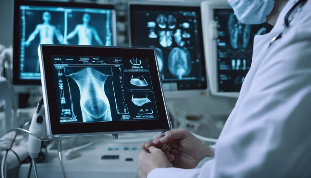
In managing bladder dysfunction, you'll find that understanding the causes is key to preventing further complications.
You should adhere to clinical evaluation protocols that recommend regular ultrasound assessments to detect issues like urinary retention early.
Bladder Dysfunction Causes
While various factors contribute to bladder dysfunction, one common cause is bladder outlet obstruction, which over time leads to increased residual volume and bladder wall hypertrophy due to chronic pressure. This condition often manifests as urinary retention, a troubling scenario where you can't completely empty your bladder.
Regularly using an ultrasound bladder scan can help monitor this residual volume effectively, offering a clear picture of the dysfunction's progression.
Further complicating matters, neurological conditions like multiple sclerosis or cauda equina syndrome severely disrupt your bladder's normal control mechanisms. This disarray can flip your life upside down, leading either to unexpected leakage or an inability to urinate despite the urgency.
Inflammation from urinary tract infections adds another layer of distress, intensifying symptoms by irritating the bladder. Such infections not only aggravate the urgency and frequency but also risk escalating into more severe complications if not addressed promptly.
Clinical guidelines stress the importance of a methodical evaluation, including a detailed assessment of symptoms, physical examination, and fundamentally, imaging studies like ultrasound.
These steps are essential to tailoring your treatment effectively, aiming to restore bladder function and prevent further complications, ensuring you regain control and improve your quality of life.
UTI Prevention Strategies
Understanding the causes of bladder dysfunction underscores the need for effective preventive measures against urinary tract infections (UTIs), particularly in medical settings. A key strategy lies in monitoring bladder volume accurately to avoid unnecessary catheterization, which directly correlates with a higher risk of catheter-associated urinary tract infections (CAUTIs).
You can greatly minimize these risks by integrating non-invasive methods such as portable ultrasound scanners to measure post-void residual volume (PVR). This approach not only helps in determining the actual necessity for catheterization but also aids in timely intervention, thereby helping to prevent infection. The Healthcare Infection Control Practices Advisory Committee (HICPAC) strongly recommends this practice as part of routine care.
Moreover, bolstering education for healthcare workers about the indications for and risks associated with catheter use is essential. A well-informed staff is more likely to adhere to guidelines that advocate for minimal catheter use and engage in vigilant management of those already catheterized.
This dual approach of using ultrasound technology for accurate measurement and enhancing staff training forms a robust defense against UTIs, particularly CAUTIs, improving patient outcomes greatly.
Clinical Evaluation Protocols
You'll find that evaluating bladder volume through ultrasound is a cornerstone in the clinical evaluation of urinary retention and bladder dysfunction. This non-invasive technique lets you accurately measure post-void residual (PVR) volume, critical for diagnosing conditions like urinary retention and bladder outlet obstruction.
The American Urological Association (AUA) recommends using bladder scanners or ultrasound within 10 minutes post-voiding to guarantee precise PVR measurements. Normally, values should be less than 50 mL.
Using a 2-5 MHz curvilinear probe placed just above the symphysis pubis, ultrasound measurements calculate bladder volume with the ellipsoid formula: depth x width x height x 0.52. It's essential you perform these assessments regularly to manage urinary conditions effectively; elevated PVR can signify risks for urinary tract infections (UTIs) and other complications if overlooked.
Clinical guidelines stress the importance of systematic evaluations. These include taking a detailed patient history, conducting thorough physical examinations, and utilizing imaging studies.
Together, these elements guide evidence-based management strategies for urinary retention and bladder dysfunction, guaranteeing you're providing ideal care based on the latest clinical standards.
Educational Resources and Updates
Signing up for POCUS 101 updates offers healthcare professionals essential educational content and resources on ultrasound techniques and bladder volume assessment. These resources are crafted to boost your scanning skills, greatly enhancing diagnostic accuracy and optimizing patient care. You'll stay on the cutting edge of technological advancements and best practices for measuring bladder volume.
Here's what you can look forward to in each update:
| Feature | Benefits | Impact on Practice |
|---|---|---|
| Latest Advancements | Guarantees you're equipped with the newest tools and methods. | Keeps your practice modern and effective. |
| Calculation Methods | Access to easy-to-use online calculators. | Speeds up your clinical decision-making. |
| Professional Development | Continuous learning opportunities. | Sharpens your skills, boosts confidence. |
| Case Studies | Real-world applications of ultrasound techniques. | Enhances understanding, improves outcomes. |
These updates not only inform you about effective bladder volume assessment methodologies but also encourage ongoing professional development. You'll find that using the provided tools, like online calculators and user-friendly interfaces, allows for quick and accurate bladder volume estimation. Keep up with POCUS 101 and guarantee your practice remains at the forefront of clinical excellence.
What are the Benefits of Assessing Bladder Volume Using Ultrasound?
Assessing bladder volume using an ultrasound scanner for bladder accuracy offers numerous benefits. It allows for non-invasive and real-time measurements, leading to more accurate diagnoses and treatment plans. Additionally, it reduces the risk of patient discomfort and eliminates the need for invasive catheterization procedures.
Frequently Asked Questions
How to Measure Bladder Volume on Ultrasound?
To measure your bladder volume on ultrasound, you'll first undergo patient preparation, ensuring your bladder is adequately filled.
An ultrasound technician will use imaging modalities, focusing on bladder anatomy through techniques like the ellipsoid formula. This method assesses the width, depth, and height of your bladder.
Understanding these ultrasound techniques is essential, as they help diagnose various bladder pathologies and have significant clinical implications for conditions like urinary retention.
How Accurate Is the Bladder Volume on Ultrasound?
You're likely wondering about the accuracy of bladder volume measured by ultrasound.
Despite various imaging techniques and frequency selections, errors can be up to 25%. Proper patient preparation is essential for minimizing these discrepancies.
Factors like bladder distension effects and wall characteristics also play significant roles.
Always assess the clinical significance of the measurements, combining them with other evaluations to guarantee accuracy.
It's a useful tool but requires careful interpretation.
How Do You Calculate Volume on Ultrasound?
To calculate volume on ultrasound, you'll need to understand the principles behind ultrasound imaging and learn specific measurement techniques.
Start by preparing the patient correctly, ensuring clear visibility of the bladder anatomy to avoid imaging artifacts.
Apply the ellipsoid formula—depth multiplied by width and height, adjusted by a coefficient—to the measurements obtained from the ultrasound.
This method is essential in clinical applications like monitoring urinary retention.
What Is the Normal Volume of the Urinary Bladder in Ultrasound?
You'd typically find that normal bladder capacity on ultrasound imaging falls below 500 mL.
Measurement techniques consider age factors, impacting normal values, which range from 300-400 mL for comfort.
In clinical significance, ensuring accurate bladder volume assessments is essential for diagnosing conditions related to urinary retention and bladder function.
Conclusion
You've learned how to assess bladder volume with ultrasound, mastering the POCUS technique and understanding its clinical relevance. From positioning the probe to applying the ellipsoid formula, you're equipped to evaluate post-void residuals accurately. Keep up with the latest guidelines and refine your skills through ongoing education. Remember, proper coding and compliance will guarantee you're not only effective but also reimbursed for your efforts. Keep practicing, and you'll enhance patient care in no time.
