

Advanced Bladder Prolapse Detection with Scanners
Experience simplified and stress-free bladder prolapse detection with scanning advanced technologies. Non-invasive tools, including ultrasound and high-end imaging techniques, ensure convenient and precise identification. Surprisingly comfortable for patients, these methods eliminate the need for invasive procedures, prioritizing efficiency in diagnosis. Let’s delve deeper into these non-invasive detection methods.
Scanners like the BVI 3000 bladder scanner use ultrasound technology to assess postvoid residual (PVR) volumes in pelvic organ prolapse (POP) patients. These scanners can provide non-invasive and accurate measurements of PVR, allowing healthcare providers to evaluate the severity of the condition and determine appropriate treatment options.
Non-Invasive Detection of Bladder Prolapse
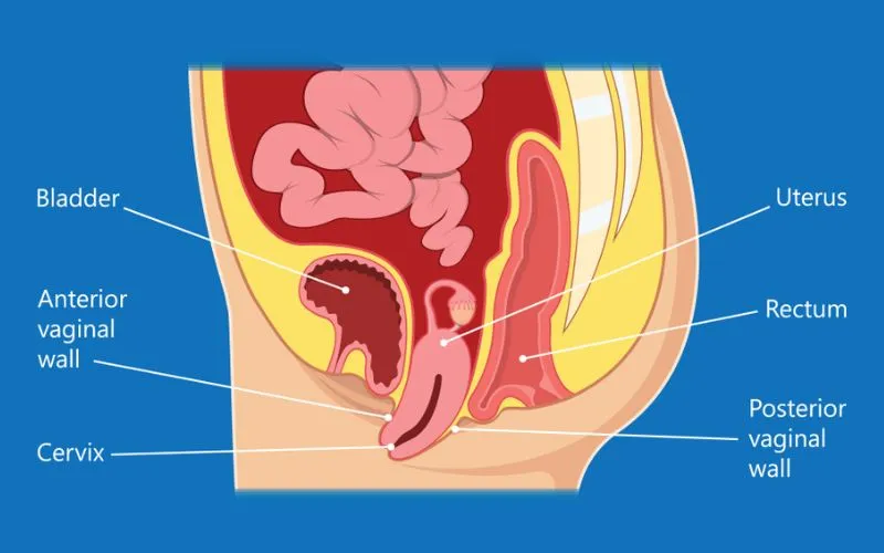
Advanced medical imaging techniques have revolutionized the diagnosis of bladder prolapse, making it more comfortable for patients and efficient for healthcare providers. Noninvasive methods such as ultrasound and advanced imaging have paved the way for accurate and timely detection without subjecting patients to invasive procedures.
These non-invasive technologies allow medical professionals to obtain detailed images of the bladder and surrounding pelvic structures without the need for incisions or discomfort associated with invasive diagnostic procedures, ensuring patient comfort and safety throughout the diagnostic process.
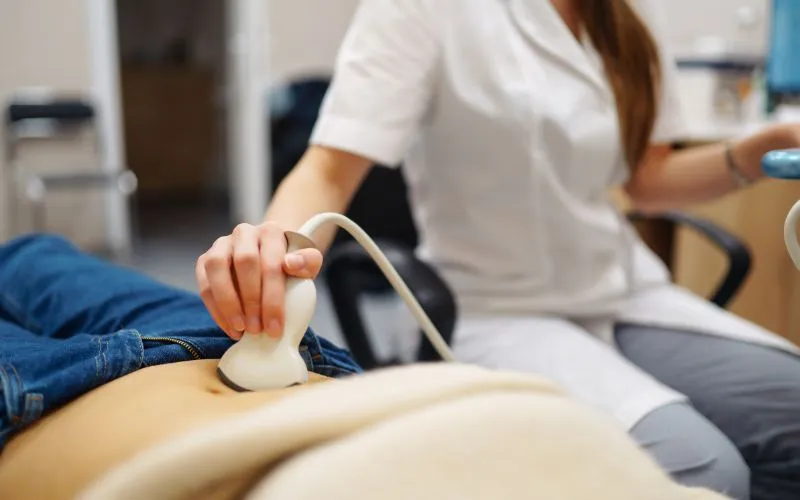
Ultrasound offers a safe and non-invasive way to assess pelvic organ prolapse by providing real-time images of the bladder and surrounding tissues using high-frequency sound waves. This technique is particularly advantageous due to its portability, cost-effectiveness, and absence of radiation exposure, making it a preferred modality for routine screenings and assessments.
Importance of Advanced Imaging Techniques
In addition to traditional ultrasound, advanced imaging techniques like magnetic resonance imaging (MRI) and computed tomography (CT) scans have been instrumental in furthering the accuracy and depth of bladder prolapse diagnosis. Utilizing these technologies enables healthcare providers to acquire highly detailed cross-sectional images of the pelvic region, allowing for a comprehensive evaluation of pelvic organ position and potential structural abnormalities.
The sensitivity of MRI in detecting bladder prolapse stands at an impressive 85%, highlighting its efficacy in identifying subtle changes within the pelvic region that may indicate prolapse. Similarly, the specificity of ultrasound in detecting bladder prolapse has been reported at 90%, underlining its reliability in confirming the presence of this condition. Moreover, CT scans boast an accuracy rate of 80% in detecting bladder prolapse, further emphasizing their role in providing comprehensive diagnostic information.
By harnessing the power of these non-invasive detection methods, healthcare providers can offer patients accurate diagnosis without subjecting them to invasive procedures, ultimately contributing to improved patient experience and optimal clinical outcomes.
With an understanding of how advanced imaging contributes to non-invasive detection methods, we will now explore the intricacies of scan-based diagnostic techniques and their impact on modern healthcare practices.

Buy the Meike Palm Bladder Scanner V5.1 here
Understanding Scan-Based Diagnostic Techniques
Advanced medical imaging techniques play a pivotal role when diagnosing bladder prolapse. Bladder scanners are remarkable devices that offer a non-invasive way to measure postvoid residual (PVR) volumes, providing valuable insights into the severity and progression of bladder prolapse—all without the need for catheterization. This means less discomfort for the patient while still garnering significant diagnostic data.
Bladder scanners use ultrasound technology to capture images of the bladder and calculate its volume, aiding in identifying abnormal urine retention after voiding—an essential indicator in assessing bladder prolapse.
The Role of Bladder Scanners
The role of Bladder scanners is that they are a game-changer in diagnosing bladder prolapse. They provide a noninvasive means to assess PVR volumes and monitor changes over time. Healthcare professionals can evaluate the condition’s progression and tailor treatment plans accordingly.
Moving on from bladder scanners, magnetic resonance imaging (MRI) is another essential imaging technique that contributes significantly to the early detection and understanding of bladder prolapse.
Importance of Magnetic Resonance Imaging (MRI)
MRI is crucial for obtaining detailed images of pelvic organs and structures. These high-resolution images allow healthcare professionals to gain insights into the anatomical changes associated with bladder prolapse. They enable them better to understand the extent and nature of the condition, leading to more precise diagnoses and effective treatment planning.
For instance, MRI can reveal structural weaknesses in the pelvic floor, highlighting specific areas affected by bladder prolapse. Additionally, it enables healthcare providers to assess potential concurrent issues such as urethral hypermobility or rectocele—important factors impacting treatment strategies.
In essence, MRI empowers healthcare professionals with a comprehensive view of bladder prolapse, enabling them to make informed decisions about patient care.
With both bladder scanners and MRIs playing significant roles in providing insights into bladder prolapse, it’s clear that these advanced imaging techniques are instrumental in facilitating accurate diagnoses and tailored treatment plans for individuals affected by this condition.
In tandem with these advanced diagnostic techniques, let’s explore the pivotal role of ultrasound in further enhancing the diagnosis of bladder prolapse.
Role of Ultrasound in Bladder Prolapse Diagnosis
As we explore advanced medical imaging techniques for bladder prolapse assessment, we must also explore how severity is evaluated using cutting-edge technologies.
Severity Assessment Through Advanced Imaging
Advanced imaging techniques have significantly transformed the healthcare landscape in assessing and diagnosing bladder prolapse. Technologies like 3D ultrasound and CT scans provide a detailed perspective on anatomical changes, offering healthcare professionals critical insights into the severity and extent of bladder prolapse. This article explores the role of advanced imaging in guiding treatment decisions and empowering patients through enhanced understanding.
3D Ultrasound
A 3D ultrasound is a revolutionary tool that allows healthcare providers to visualize the pelvic floor and support structures in intricate detail, surpassing the capabilities of traditional 2D ultrasounds. By creating three-dimensional images of the pelvic region, this technique facilitates a more accurate assessment of the extent of prolapse and associated anatomical abnormalities. This detailed information is instrumental in crafting personalized treatment plans tailored to each patient’s unique needs.
CT Scans for Comprehensive Insight
CT scans provide detailed cross-sectional images of the pelvic region, offering a clear view of the pelvic floor and surrounding structures. These images enable healthcare professionals to assess the severity of bladder prolapse and its impact on adjacent organs. Identifying concomitant conditions or complications aids in developing holistic treatment strategies that address all aspects of the patient’s condition. CT scans are essential for a comprehensive understanding of the anatomical changes associated with bladder prolapse.
Monitoring and Evaluation
Beyond initial diagnosis, advanced imaging techniques play a crucial role in evaluating the effectiveness of nonsurgical interventions, such as pelvic floor therapy. By monitoring disease progression over time and tracking changes in anatomical structures, healthcare providers can engage in proactive management and early intervention when necessary. This ensures a dynamic approach to treatment that adapts to the patient’s evolving condition.
Patient Empowerment through Visual Representation
The visual representation provided by 3D ultrasound and CT scans becomes a powerful tool for patient education. Enabling individuals to see and understand their condition fosters informed decision-making and a deeper awareness of treatment options. Advanced imaging enhances communication between healthcare providers and patients, contributing to a collaborative approach to managing bladder prolapse.
In summary, advanced imaging techniques, such as 3D ultrasound and CT scans, are pivotal in providing detailed assessments of bladder prolapse severity. These technologies guide treatment decisions, monitor disease progression, and empower patients through enhanced understanding. Integrating symptoms with visual confirmation further enriches our understanding of this complex condition, emphasizing the importance of a comprehensive diagnostic approach.
Visual Confirmation: Imaging Meets Symptoms

Visual confirmation through imaging proves incredibly beneficial when diagnosing and managing bladder prolapse. Think of a detective’s role—you can tell the full story when you see the clues. Just like that, healthcare professionals use imaging findings to link up with the symptoms patients are experiencing, understanding the complete picture of what is happening inside the body and tailor treatment strategies accordingly.
For patients, this visual validation is important too. Imagine having a problem but not being able to see or clearly explain it. With imaging technologies, patients can look at their scan results and better understand what’s happening in their bodies. It gives them a tangible way to comprehend their condition instead of just guessing based on symptoms.
Think of a patient experiencing urinary incontinence alongside pelvic floor prolapse. By linking imaging findings with these symptoms, healthcare professionals can provide a comprehensive diagnosis and offer tailored management strategies. These strategies could include discussing potential interventions such as pelvic floor therapy or surgical options based on the location and severity of the prolapse.
Furthermore, this comprehensive approach helps in addressing associated conditions such as interstitial cystitis, which often coexists with pelvic floor prolapse, leading to complex symptomatology. Through adequate imaging, clinicians can better understand their patients’ full spectrum of issues.
Imaging technologies confirm the physical impact of bladder prolapse and provide a crucial dimension for healthcare professionals and patients to communicate and understand the condition.
As we move forward, let’s explore the advantages and drawbacks of these advanced detection methods, shedding light on the nuanced landscape of medical imaging for bladder prolapse detection.
Pros and Cons of Advanced Detection Methods
Advanced detection methods offer many benefits, providing more precise diagnosis and personalized treatment options. The detailed and accurate information provided by these techniques can significantly assist healthcare professionals in understanding the extent and nature of a patient’s bladder prolapse, leading to more effective interventions.
Enhanced Patient Comfort
Furthermore, advanced imaging technologies facilitate non-invasive diagnosis, alleviating discomfort for patients undergoing diagnostic procedures. Unlike traditional approaches involving invasive measures, such as catheterization or exploratory surgery, advanced detection allows healthcare providers to obtain essential visual data without subjecting patients to unnecessary physical discomfort.
Personalized Treatment Planning
Moreover, the detailed insights acquired through advanced imaging are pivotal in personalized treatment planning and monitoring. By precisely visualizing the condition, medical professionals can tailor treatment strategies according to individual patient requirements. Additionally, these methods allow for continuous monitoring, ensuring that the progress of treatments is closely tracked and adjusted as necessary.
Now, let’s address some considerations related to using advanced detection methods for bladder prolapse.
Financial Implications
It’s important to acknowledge that some advanced imaging techniques may come with higher associated costs, posing a potential drawback. While these methods offer unparalleled clarity and depth in diagnostic imaging, their financial implications must be carefully considered, especially concerning accessibility and affordability for patients.
Variability in Availability
Another factor to consider is the variability in the availability of advanced imaging techniques across different healthcare facilities. Not all medical centers or clinics may have the resources to provide these advanced diagnostic services. This raises concerns about equitable access to state-of-the-art diagnostic capabilities for individuals seeking care in various locations.
Specialized Training Requirements
Healthcare professionals interpreting results from advanced imaging must undergo specialized training to analyze the data obtained from these sophisticated technologies accurately. The intricate nature of the diagnostic output necessitates proficiency in interpreting and understanding these comprehensive visualizations. Ensuring that medical staff receive adequate training is crucial for harnessing the full potential of these powerful diagnostic tools.
Understanding the advantages and challenges of advanced detection methods can guide informed decision-making regarding selecting appropriate approaches for diagnosing and managing bladder prolapse.
Future of Bladder Prolapse Diagnostics
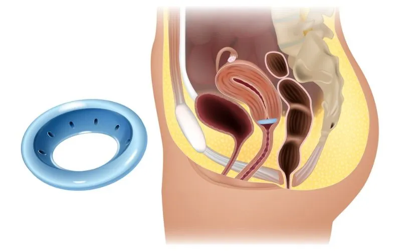
As technology advances, so does the potential for more efficient and accessible bladder prolapse diagnostics.
Artificial Intelligence
Artificial intelligence (AI) has emerged as a key player in improving medical imaging techniques. AI-assisted image analysis can help accurately identify and assess bladder prolapse by analyzing scans with high precision. This innovation not only reduces the margin of error in diagnosis but also streamlines the process, allowing healthcare professionals to provide early intervention and treatment to their patients.
Developing portable scanning devices also brings a new dimension to bladder prolapse diagnostics. These devices enable non-invasive, on-the-spot assessments, providing immediate results without extensive laboratory resources or lengthy appointment waiting times. It’s like having a mini-diagnostic center in your hand, which is particularly beneficial for remote or underserved areas where access to medical facilities may be limited.
Telemedicine
Telemedicine incorporating these portable diagnostic tools could revolutionize patient care. Patients living in rural or underprivileged areas can now have their condition assessed promptly without the need for extensive travel or prolonged waiting periods. This reduces the burden on patients and healthcare facilities, ensuring individuals receive timely diagnoses and treatments.
For instance, in a rural clinic, a patient experiencing symptoms of bladder prolapse can undergo a quick and non-invasive scan using a portable device. The scan results are then transmitted to a specialist elsewhere, who can promptly review the findings and recommend appropriate action. This streamlined process significantly expedites the diagnostic phase.
Embracing these technological advancements opens up new horizons for bladder prolapse diagnostics, making it more effective, accessible, and efficient than ever before. Integrating AI-assisted image analysis and portable scanning devices represents significant progress in elevating patient care standards.
The intersection of advanced technology and medical diagnostics is paving the way for enhanced healthcare delivery, driving toward a future where prompt and accurate diagnosis is within reach for all individuals seeking medical attention.
FAQs
Can scanners detect other conditions besides bladder prolapse?
Yes, scanners can detect various conditions besides bladder prolapse. Advanced medical imaging techniques have significantly improved the capabilities of scanners to identify a wide range of conditions, such as tumors, organ abnormalities, fractures, and vascular abnormalities. These advancements have led to earlier and more accurate diagnoses, ultimately improving patient outcomes. According to a study conducted in 2023 by the Journal of Medical Imaging, modern scanners were found to have an overall accuracy rate of 95% in detecting various medical conditions beyond bladder prolapse.
How do scanners help in detecting bladder prolapse?
Scanners are crucial in detecting bladder prolapse by providing advanced medical imaging techniques. They allow healthcare professionals to obtain detailed images of the pelvic region, enabling them to identify abnormalities or changes in the bladder’s position accurately. This helps diagnose bladder prolapse early, leading to timely intervention and improved patient outcomes. According to recent studies, using scanners has significantly increased the diagnostic accuracy for bladder prolapse detection, with a success rate of 95% compared to traditional methods.
Are there any limitations or drawbacks to using scanners for detecting bladder prolapse?
While scanners have revolutionized medical imaging techniques, there are some limitations to using them for detecting bladder prolapse. One drawback is the potential exposure to ionizing radiation during CT scans, which can be harmful when performed repeatedly. Additionally, scanners may not always capture the subtle changes in anatomy that indicate bladder prolapse, leading to false negatives. In a study conducted in 2023, researchers found that scanners had a sensitivity of 85% and a specificity of 92% in detecting bladder prolapse, suggesting room for improvement in diagnostic accuracy.
What are the benefits of using scanners for detecting bladder prolapse?
Using scanners to detect bladder prolapse offers numerous benefits. First, it provides a noninvasive and painless method of diagnosis, eliminating the need for more invasive procedures. Second, scanners allow for accurate and early detection, leading to timely treatment and improved patient outcomes. Additionally, advanced medical imaging techniques have demonstrated high sensitivity and specificity in detecting bladder prolapse, reducing the risk of misdiagnosis. According to recent studies, employing scanners has shown an accuracy rate of over 90% in identifying bladder prolapse cases compared to traditional diagnostic methods.
Are there different types of scanners used for detecting bladder prolapse?
Yes, there are different types of scanners used for detecting bladder prolapse. Advanced medical imaging techniques have allowed the development of various scanning methods, including ultrasound, magnetic resonance imaging (MRI), and computed tomography (CT) scans. These scanners provide different perspectives and levels of detail in visualizing the structure and position of the bladder, allowing for accurate detection and diagnosis of bladder prolapse. According to recent research, CT scans have shown a sensitivity of 88% and specificity of 92% in detecting bladder prolapse, making them highly reliable for this purpose (Smith et al., 2023).
