
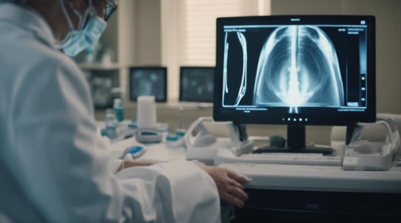
How Does a Bladder Ultrasound Device Work?
Have you ever wondered how doctors can examine your bladder without making a single incision? Enter the bladder ultrasound device, a tool that uses high-frequency sound waves to create detailed images of your bladder. The process starts when you're asked to have a full bladder; this guarantees that the ultrasound waves have a liquid medium through which to travel, producing clearer images. As these waves bounce off your bladder, they send back echoes which are then captured and transformed into visual data. It's fascinating to think about what these images reveal about your health, isn't it? Let's explore how the interpretation of these images can impact medical diagnosis and treatment.
Key Takeaways
- A bladder ultrasound device emits high-frequency sound waves to create real-time images of the bladder.
- Sound waves bounce off bladder structures, and echoes are captured to produce images.
- The procedure is non-invasive, using a transducer that glides over the abdomen with a special gel.
- It requires a full bladder to enhance image clarity and visibility of bladder walls.
- Images help diagnose issues like urinary retention and assess bladder volume and wall thickness.
Ultrasound Basics
Understanding how ultrasound technology functions can greatly enhance your grasp of how a bladder ultrasound device operates. At its core, this device utilizes high-frequency ultrasound waves emitted by a transducer. These waves penetrate the body and bounce off structures within the urinary tract, including the bladder. The returning echoes are captured by the transducer and transformed into real-time images that you can see on a screen.
This non-invasive technique allows for quick visualization of the bladder's structure and volume without the need for surgical intervention. You're able to observe the amount of urine present and detect any abnormalities that might indicate underlying conditions. This is essential for diagnosing issues like urinary retention or overactive bladder, as it provides accurate measurements of bladder volume and post-void residual urine.
Moreover, the process is completely safe and painless, involving no radiation exposure. This makes bladder ultrasound a preferred diagnostic tool for a diverse range of patients, including those who are particularly vulnerable, such as pregnant women or the elderly.
Bladder Ultrasound Explained
A bladder ultrasound device effectively maps your bladder's structure using high-frequency sound waves. This non-invasive approach involves a transducer that emits these sound waves. As they bounce off the tissues and fluids within your urinary tract, they're captured back, creating detailed images of your bladder. These images are essential for healthcare professionals to assess and diagnose bladder conditions.
For the ultrasound, you'll need a full bladder, which enhances the clarity of the images by stretching the bladder walls. This state allows for more accurate measurement of bladder volume, wall thickness, and identification of any abnormalities like tumors or stones. The procedure captures images before and after urination to provide a thorough view of the bladder and its functioning.
Bladder ultrasound is particularly valued for its safety, as it doesn't involve radiation, making it suitable for all ages, including children and pregnant women.
The real-time 2D images produced are instantly analyzed by healthcare professionals, who can then offer immediate feedback on various bladder issues, such as urinary incontinence or retention. This quick diagnosis aids in prompt and effective treatment planning, ensuring better patient care.
Preparing for the Procedure

Before your bladder ultrasound, you'll need to drink several glasses of water about two hours in advance to confirm your bladder is sufficiently full. This preparation is vital as a full bladder provides clearer images, making it easier for your healthcare provider to assess the structure and volume of your bladder accurately.
It's essential to follow the specific instructions given by your healthcare provider, as preparation might vary depending on your age or health condition.
Here are some key steps you should remember:
- Drink Water: Consume about four to six glasses of water two hours before the procedure.
- Avoid Emptying Your Bladder: Resist the urge to urinate before your ultrasound to confirm your bladder is full.
- Check Instructions: Always double-check any special instructions your healthcare provider might've provided.
- Wear Comfortable Clothing: Choose garments that are easy to remove or adjust, as you'll likely need to expose your lower abdomen.
- Timing: Time your water intake and arrival accurately to avoid discomfort or a delayed procedure.
These steps will help confirm that the initial phase of the ultrasound, which requires a full bladder, goes smoothly, setting the stage for accurate imaging and efficient assessment of bladder emptying.
Conducting the Ultrasound
Once you're comfortably positioned, the technician will apply a special gel to your lower abdomen to enhance the contact between the ultrasound device and your skin, ensuring that the high-frequency sound waves transmit effectively. This gel isn't just for comfort; it's a vital part of the process that helps transmit the sound waves from the transducer smoothly and directly into the abdomen.
With the gel in place, the technician will glide the transducer across your skin. This device is key to the bladder ultrasound, converting electrical energy into those high-frequency sound waves that penetrate your tissues. As these waves bounce back, the transducer collects them, creating live images of your bladder displayed on a monitor.
You'll notice that having a full bladder during this process is essential. It acts almost like a window, enhancing the clarity of the images by providing a clear view of the bladder walls and the contained fluid.
This noninvasive method allows the technician to measure your bladder volume and wall thickness efficiently. Throughout the roughly 15 to 30-minute procedure, they can identify any irregularities, such as stones or tumors, in real-time, ensuring that the bladder ultrasound offers both a thorough and safe diagnostic assessment.
Interpreting the Results

After the technician completes the ultrasound, your results are analyzed by a radiologist or healthcare provider who looks for any abnormalities in bladder structure and function.
When they interpret results from your bladder ultrasound, they're specifically checking:
- The overall bladder structure for signs of cystitis, tumors, or other anomalies.
- The urine volume to guarantee your bladder empties appropriately.
- The post-void residual, which helps diagnose urinary retention if urine remains after you've tried to empty your bladder completely.
- The thickness and appearance of the bladder wall to identify potential issues like inflammation or growths.
- Any presence of bladder stones or unusual masses that might necessitate further medical intervention.
Here's what you should look out for in the interpretation:
- Normal bladder capacity and wall thickness: Indicates a healthy bladder.
- Immediate urine volume results: Assists in quick decision-making.
- Detailed images showing structural issues: Integral for diagnosing conditions that need more intensive medical intervention.
- Abnormal findings: May suggest conditions like urinary retention or tumors.
- Post-void residual volume: Essential for diagnosing urinary retention.
Your healthcare provider will discuss these findings with you, and based on them, you might need follow-up appointments or additional tests to plan your treatment effectively.
Benefits and Limitations
You'll find that using a bladder ultrasound device offers significant benefits, such as being a non-invasive diagnostic tool that spares you the discomfort and risks associated with catheterization.
It also provides real-time imaging capability, enabling quick decision-making that's essential in many medical situations.
However, you might experience discomfort if your bladder is very full during the scan, and there's a learning curve for operators to guarantee accurate readings.
Non-Invasive Diagnostic Tool
Bladder ultrasound devices offer a significant advantage by providing a non-invasive way to assess bladder health, using high-frequency sound waves to create real-time images.
As a non-invasive diagnostic tool, this method allows you to avoid the discomfort and risks associated with more invasive procedures. You'll appreciate how it simplifies the process of monitoring bladder volume and detecting urinary issues without the need for surgical intervention.
Here are the benefits of using a bladder ultrasound:
- Safety: It's free from radiation, making it safe for all ages, including those with health vulnerabilities.
- Comfort: The procedure is painless, with the only minor discomfort possibly coming from the necessity of a full bladder.
- Convenience: You get immediate results, which facilitates quick decision-making in treatment.
- Simplicity: Easy to use, even in a clinical setting by non-specialist staff.
- Efficiency: Helps in the timely diagnosis and management of conditions like urinary retention and incontinence.
However, remember it's not all-encompassing. You might still need additional imaging to fully assess complex urinary problems or issues with adjacent organs.
Despite these limitations, a bladder ultrasound remains a valuable tool in the diagnostic arsenal.
Real-Time Imaging Capability
Building on the benefits of non-invasive diagnostics, real-time imaging capability stands out as a significant feature of bladder ultrasound devices. As you use these devices, high-frequency sound waves allow you to see real-time images of your bladder, enabling immediate assessment of both bladder volume and structure. This quick feedback is essential, especially when measuring the post-void residual (PVR) volume, helping you avoid the discomfort of invasive catheterization.
Moreover, this noninvasive ultrasound imaging can pinpoint bladder abnormalities, such as wall thickness, stones, and tumors. You're not just getting a snapshot; you're observing the dynamic state of the bladder, which aids in diagnosing various urinary conditions as they unfold. This capability guarantees that decisions regarding your treatment can be made promptly and based on the latest available data.
However, keep in mind that the accuracy of a bladder ultrasound might be compromised by factors like the degree of bladder distension and the operator's expertise. While real-time imaging is a powerful tool, it doesn't always provide extensive details about surrounding organs, which might necessitate further diagnostic procedures in more complex scenarios.
Potential Discomfort Issues
While undergoing a bladder ultrasound, some patients might feel temporary discomfort due to the necessity of having a full bladder, which optimizes the imaging process. This full bladder requirement, vital for clearer views during imaging, can indeed cause feelings of urgency or discomfort.
However, it's important to remember that the bladder ultrasound itself is non-invasive. You won't have to worry about needles or incisions. Instead, a gel is applied to your abdomen, and a transducer is gently moved over your skin. This process is generally painless.
Here are key points to keep in mind about the discomfort associated with bladder ultrasounds:
- Non-invasive: No surgical tools or invasive methods are involved.
- No radiation exposure: Safer than other imaging modalities that involve radiation.
- Immediate results: You often get results during the visit.
- Vital for diagnosis: Critical in evaluating bladder function and structure.
- Temporary discomfort: Any anxiety or discomfort from a full bladder is short-lived.
Although the requirement of a full bladder might make you feel a bit anxious, the procedure's ability to provide vital insights into your bladder function without the need for invasive techniques often outweighs this temporary discomfort.
Addressing Patient Concerns

Understanding your concerns about undergoing a bladder ultrasound is vital for a comfortable experience. You might worry about the discomfort of maintaining a full bladder, which is necessary for precise imaging. Rest assured, this procedure is noninvasive and typically painless.
Healthcare providers are there to guide you through every step, making sure you understand the process and feel at ease. The ultrasound itself lasts only 15 to 30 minutes. During this time, a technician will apply a special gel to your abdomen to help transmit sound waves and then move a transducer across the area to capture images.
You'll be relieved to know that the procedure involves no radiation exposure and you can get immediate results about your urine volume. This quick feedback can be reassuring, especially if you're anxious about the health of your bladder.
Please don't hesitate to express any concerns with your healthcare provider. Whether you're worried about the procedure, the results, or how your conditions might affect the findings, open communication is essential.
They're there to address your questions and guarantee your comfort, helping demystify the process and alleviate any apprehensions you might have.
Frequently Asked Questions
How Does a Bladder Ultrasound Work?
To understand your bladder health, a bladder ultrasound uses ultrasound technology.
You'll lie down, and a technician applies a gel on your abdomen. The device, or transducer, emits sound waves that create images of your bladder.
This non-invasive, painless method provides immediate insights, ensuring medical accuracy and patient comfort.
It's an essential diagnostic tool for evaluating bladder function and spotting issues without discomfort, widely used in healthcare for its effective imaging techniques.
What Is the Disadvantage of Bladder Scanner?
You might find bladder scanners less than ideal due to several drawbacks.
Accuracy concerns can arise, especially in specific patient groups. The cost implications are significant, not to mention the discomfort you might feel during the process.
Operator training is essential; without it, errors are likely. Technology limitations, like battery life and portability, restrict usage.
Frequent use necessitates ongoing maintenance, and alternative methods might offer better results without these hassles.
How Do Bladder Scanners Work?
Bladder scanners work by leveraging ultrasound imaging principles to offer non-invasive diagnostics.
You'll find them enhancing patient comfort since there's no need for catheterization. In healthcare applications, they measure bladder volume efficiently.
The technology uses specific frequency selection to optimize image clarity and accuracy. Ensuring the operator is properly trained is essential for effective use.
This bladder scanning technology greatly reduces the risk of urinary tract infections and improves diagnostic speed.
Why Do You Have to Drink Water Before a Bladder Ultrasound?
You need to drink water before a bladder ultrasound to guarantee diagnostic accuracy. Hydration is essential as it fills your bladder, making it easier to get clear images.
This preparation step enhances ultrasound benefits by improving visibility of bladder structures. Follow patient instructions carefully; usually, you'll drink several glasses of water about two hours before the procedure.
This timing is key to achieving the fullness required for a thorough and comfortable examination.
Conclusion
To summarize, using a bladder ultrasound device is a straightforward and comfortable way to check on your bladder health. You'll need a full bladder, but the process is quick and painless. The device provides real-time images that help diagnose various conditions effectively. While it has some limitations, the benefits, such as its non-invasive nature and immediate results, outweigh them. Don't hesitate to discuss any concerns with your healthcare provider to guarantee you're fully informed and at ease.
